
Bone grafting is a predictable surgical technique used to rebuild and strengthen the jaw when natural bone has been lost. For many patients, grafting isn’t just about supporting a single tooth — it preserves the contours of the jaw, supports adjacent teeth, and maintains the proportions of the face. When bone volume declines, patients can experience changes in chewing ability, shifting teeth, and a sunken appearance that affects facial balance.
Loss of bone can make routine dental care more complicated and may limit options for long-term tooth replacement. A graft restores a stable foundation so restorative treatments — particularly dental implants — have the bone mass they require. In short, a successful graft gives patients more predictable functional and cosmetic results than attempting restorations on a compromised ridge.
Because the jawbone responds to mechanical forces, maintaining its volume is also important for oral health beyond immediate repairs. Proper bone height and width help distribute chewing forces evenly, reduce stress on neighboring teeth, and contribute to long-term prosthetic success. That functional stability is one reason many clinicians recommend grafting as a proactive step when bone loss is evident.
Several common conditions lead to bone loss in the jaws. Tooth extraction without socket preservation, advanced periodontal (gum) disease, trauma from accidents, and long-term tooth loss each remove the stimulation that keeps bone healthy. When the tooth root is absent, the body resorbs bone in that area because the mechanical signals that sustain it are reduced.
Clinically, patients may notice changes such as denture instability, food trapping around gaps, or gradual shifting of neighboring teeth. Radiographic evaluation often reveals reduced ridge height and width that might not be obvious from an external examination. Left unaddressed, these changes can complicate restorative plans and increase the likelihood of further dental problems.
Bone loss in the posterior maxilla (upper jaw behind the molars) presents a particular challenge because of the proximity to the sinus cavity. In these cases, insufficient vertical bone height can prevent implant placement unless additional bone is added beneath the sinus floor. Understanding the cause and pattern of bone loss helps the clinician develop a targeted grafting strategy tailored to each patient's anatomy and future treatment goals.
Graft materials fall into several categories, and the choice depends on clinical needs, patient preferences, and the desired biological response. Autografts use the patient’s own bone — typically harvested from the jaw, chin, or hip — and offer living cells and growth factors that actively contribute to new bone formation. Because the material is the patient’s own tissue, autografts are highly effective for promoting integration, though they require an additional donor site.
Allografts are sourced from human donors and undergo processing to make them safe for use. These materials act primarily as a scaffold that new bone can grow into and are commonly used when avoiding a second surgical site is preferred. Xenografts, most often derived from bovine sources, provide a long-lasting framework that slowly resorbs while host bone replaces it. Synthetic options (alloplasts) — such as calcium phosphate or bioactive glass — are manufactured to mimic bone structure and are useful when predictable, standardized graft material is desired.
Each material supports bone formation through one or more biological mechanisms: osteoconduction (providing a scaffold), osteoinduction (stimulating precursor cells to form bone), or osteogenesis (contributing living bone-producing cells). Often clinicians combine materials or use biologically active additives to enhance outcomes, tailoring the approach to the specific defect and the patient’s healing profile.
Membranes and biologic agents are frequently used alongside grafts to protect the area, guide tissue growth, and reduce soft-tissue ingrowth into the graft site. These adjuncts help create the stable environment necessary for predictable bone regeneration and can shorten the timeline to definitive restoration.
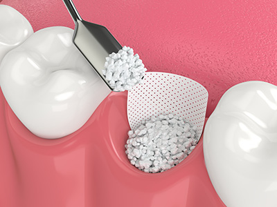
Socket preservation is a common, minimally invasive grafting procedure performed at the time of tooth extraction. Placing graft material into the socket immediately helps limit the natural shrinkage that follows extraction and preserves the ridge for future implant placement or prosthetic work. This approach reduces the need for more extensive augmentation later and preserves soft-tissue support for esthetic outcomes.
Ridge augmentation focuses on widening or building the height of a deficient alveolar ridge. This procedure can be performed prior to implant placement or simultaneously with implant surgery, depending on the clinical scenario. Ridge augmentation is especially helpful when preparing the site for fixed bridgework or when restoring natural contours for improved esthetics.
For patients lacking height in the upper posterior jaw, a sinus lift (also called sinus augmentation) creates space beneath the sinus membrane and introduces graft material to increase vertical bone. The goal is to achieve sufficient bone to support implants without encroaching on the sinus. Sinus lifts are a routine solution for many patients who would otherwise be poor candidates for implants because of insufficient height.
More complex reconstructions may combine techniques and materials to address extensive defects from periodontal disease, trauma, or long-term tooth loss. The surgical plan is customized after a thorough clinical and radiographic evaluation, often using 3D imaging to map bone volume and determine the most conservative and effective approach for predictable regeneration.
Healing after a bone graft typically involves an initial period of soft-tissue recovery followed by gradual bone remodeling over several months. Patients can expect some localized swelling, minor discomfort, and temporary dietary adjustments as the site heals. Careful postoperative instructions — including gentle oral hygiene, short-term activity modification, and follow-up visits — support predictable outcomes.
Clinical follow-up and imaging are important to monitor graft integration and to determine the optimal timing for any subsequent restorative work, such as implant placement. The graft must become well-incorporated and stable before loading with a prosthetic tooth; that integration generally takes several months, but timing varies with the graft type, the defect size, and individual healing factors.
When successful, bone grafting restores the structural foundation for long-lasting dental restorations, improves prosthetic stability, and preserves facial contours. For patients considering implants, a stable graft significantly increases the likelihood of a durable, functional result. Even when implants are not chosen, grafting can enhance the fit and comfort of removable prostheses and improve oral health by stabilizing adjacent teeth.
In experienced hands, bone grafting is a routine part of modern dental care. The procedure requires careful diagnosis, meticulous surgical technique, and attentive postoperative management, all of which contribute to predictable, esthetic, and functional outcomes that benefit patients for years to come.
At Vernon Woods Dental & Implant Center, our team combines advanced imaging, thoughtful treatment planning, and proven regenerative materials to help patients restore their oral health and confidence. If you would like to learn more about whether bone grafting is right for you, please contact us for more information.


Bone grafting is a surgical procedure that rebuilds or augments the jawbone to restore lost volume and support dental restorations. Clinicians use grafting to recreate the contours and density of bone that are lost after tooth extraction, periodontal disease, trauma or long-term tooth absence. By restoring the ridge, a graft creates a stable foundation for implants, bridges or improved denture fit.
Beyond enabling restorations, successful grafting helps preserve facial proportions and chewing function by maintaining proper ridge height and width. Maintaining bone also reduces stress on neighboring teeth and supports long-term oral health. For many patients, a graft is the difference between predictable restorative results and limited treatment options.
Jawbone loss commonly results from tooth extraction without preservation, advanced periodontal disease, traumatic injury or prolonged absence of mechanical stimulation to the socket. When a tooth root is missing, the body gradually resorbs bone because the functional signals that sustain it are reduced. Patients may not notice early changes externally, but loss often leads to denture instability, food trapping or shifting of adjacent teeth.
Diagnosing bone loss involves a clinical exam and radiographic imaging, including periapical films, panoramic views and increasingly, CBCT scans to assess volume in three dimensions. These images reveal ridge height and width and identify defects that are not visible on visual inspection alone. Accurate diagnosis helps clinicians create a targeted grafting plan matched to the patient's anatomy and restorative goals.
Graft materials include autografts (the patient’s own bone), allografts (donor human bone), xenografts (animal-derived matrices) and synthetic alloplasts (manufactured substitutes). Autografts bring living cells and growth factors and are often considered the gold standard for regeneration but require a donor site. Allografts and xenografts primarily provide a scaffold for new bone to form and avoid the morbidity of a second surgical site.
Synthetic materials offer consistent composition and eliminate biological sourcing concerns while supporting predictable osteoconduction. Each material supports regeneration through osteoconduction, osteoinduction or osteogenesis to varying degrees, and clinicians often select or combine materials to match the clinical need. Using biologic additives or growth factors can enhance cellular activity and accelerate integration in cases with compromised healing.
Socket preservation is a preventive grafting procedure performed at the time of tooth extraction to fill the socket and limit the natural bone shrinkage that follows. This minimally invasive approach helps maintain ridge dimensions and soft-tissue support, reducing the need for later augmentation. It is commonly used when future implant placement or optimal esthetics are anticipated.
Ridge augmentation is a more comprehensive procedure that rebuilds deficient ridge height or width and can be performed before or with implant placement, depending on the situation. Augmentation may use block grafts, particulate grafts, membranes or a combination to recreate natural contours. The technique is selected based on the defect size, location and the restorative plan.
A sinus lift, or sinus augmentation, increases vertical bone height in the posterior maxilla by elevating the sinus membrane and placing graft material beneath it. This procedure creates the bone volume necessary to support implants when the natural ridge is too short due to sinus pneumatization or bone loss. Sinus lifts are routine in many implant practices and can be tailored to the patient’s anatomy and the amount of augmentation required.
Success depends on careful assessment of sinus anatomy, the health of the sinus lining and the chosen graft material and technique. Complications are uncommon when performed by an experienced clinician and include membrane perforation, which can often be managed during the procedure. A well-executed sinus lift allows predictable implant placement and long-term prosthetic stability in the upper posterior jaw.
Membranes are barrier materials used to separate the grafted bone from overlying soft tissue and to guide bone regeneration by preventing soft-tissue ingrowth. They come in resorbable and nonresorbable forms, and the selection depends on the defect and the desired healing time. Proper membrane use creates a protected environment that supports undisturbed bone formation.
Biologic agents such as platelet-rich fibrin (PRF) or recombinant growth factors can be applied to enhance cellular recruitment and accelerate healing of the graft site. These additives may improve the quality and rate of new bone formation, particularly in patients with reduced regenerative capacity. Clinicians weigh benefits against clinical evidence and patient factors when incorporating biologics into a grafting protocol.
After a bone graft, patients should expect a period of soft-tissue healing followed by slower bone remodeling that can take several months to complete. Common immediate symptoms include localized swelling, mild to moderate discomfort, and temporary changes in diet and oral hygiene routines. Short-term instructions often include gentle rinsing, avoiding pressure on the surgical site, and following medication and activity guidance provided by the team.
At Vernon Woods Dental & Implant Center, follow-up appointments and imaging are scheduled to monitor graft incorporation and to determine the timing of any subsequent restorative work. Patients are advised about signs to report, such as increasing pain, fever or prolonged drainage, which may indicate a complication. Clear communication and timely follow-up are key to achieving a stable foundation for future implants or prostheses.
The interval between grafting and implant placement depends on the graft type, defect size and individual healing capacity, with common healing windows ranging from three to nine months. Autografts may integrate faster in some cases, while xenografts and certain synthetics can require longer remodeling times. Your clinician evaluates healing through clinical exams and imaging to determine the safest time to load the site.
In select situations, implants can be placed simultaneously with grafting when primary stability is achievable and the defect is minor, which can shorten overall treatment time. Conversely, larger or more complex augmentations often require staged approaches to ensure predictable bone volume before implant placement. Decisions about timing are individualized to reduce the risk of failure and to optimize long-term outcomes.
Good candidates for bone grafting are patients who are in generally good health, have controlled systemic conditions and are committed to following postoperative care. Smoking, uncontrolled diabetes and certain medications can impair healing, so clinicians evaluate and manage these risks before recommending grafting. Age alone is not usually a limiting factor, but overall biologic healing potential and oral hygiene are important considerations.
As with any surgical procedure, risks include infection, graft exposure, insufficient integration and, in the upper jaw, potential sinus complications. Careful case selection, sterile technique and appropriate postoperative management reduce these risks significantly. If complications occur, additional interventions or alternative restorative strategies may be necessary to achieve a functional outcome.
Vernon Woods Dental & Implant Center approaches bone grafting with comprehensive planning that uses clinical evaluation, CBCT imaging and individualized treatment objectives. This allows clinicians to choose the most appropriate graft material and surgical technique while anticipating anatomic challenges. Clinicians discuss the proposed sequence of care, expected healing timeline and follow-up plan with each patient before treatment begins.
On the day of surgery, local anesthesia and sedation options are used as needed to ensure patient comfort while precise graft placement and membrane management are performed. Postoperative instructions, timely reviews and imaging confirm integration and guide the transition to restorative steps such as implant placement. The practice emphasizes conservative, well-documented approaches and coordinates care across specialties when complex reconstructions are required.
Ready to schedule your first visit and to join the Vernon Woods Dental & Implant Center family?
Scheduling your visit or getting answers to your questions is simple. The helpful team at Vernon Woods Dental & Implant Center is here to assist you! We can easily help you book appointments, explain treatment details, and address any concerns you have. Connect with us however you prefer—by phone or our quick online form. Don't delay your smile goals; contact us today and experience the comfort and confidence that personalized dental care provides.
Back to top