
Oral cancer remains a significant public health concern. Each year tens of thousands of Americans receive a diagnosis involving the lips, mouth, or oropharynx, and a substantial number of those cases can be life-changing. Much of the difference between a curable and a life-threatening case comes down to one factor: detection. When abnormalities are found early, treatment options are broader and outcomes tend to be better.
Screening is a practical, low‑risk step that dental professionals can take during a regular checkup. It doesn’t require extensive preparation from the patient and can be completed as part of a routine exam. Because early lesions are often subtle and painless, many people are unaware anything is wrong until the disease is advanced; proactive screening helps close that gap.
Including oral cancer screening in your dental care routine is not only sensible for people with clear risk factors — it is an important safeguard for everyone. Regular checks create a baseline and make it easier to spot change over time, which is exactly what clinicians rely on to recognize early warning signs.
Oral cancers can arise in several locations within the mouth and throat. Dentists commonly examine the tongue (especially the sides and undersurface), the floor of the mouth, the gums, the inner cheek lining, the hard palate and the lips. The oropharynx — including the tonsils and the back of the tongue — is another frequent site, particularly for cancers linked to viral causes.
Certain lifestyle and medical factors increase the probability of developing oral cancer. Tobacco use and heavy alcohol consumption remain two of the strongest risk contributors. In recent years, infection with human papillomavirus (HPV) has been linked to a rise in cancers of the oropharynx, shifting some of the epidemiology toward younger, otherwise healthy adults. Other influences can include prolonged sun exposure to the lips, a history of head or neck radiation, chronic acid reflux, and overall nutritional status.
While statistics show differences in risk across age and sex groups, no one should assume they are immune. A careful screening approach considers the whole person — medical history, behavioral risks, and any new or persistent symptoms — rather than relying solely on demographic averages.
A standard screening is straightforward and typically begins before the visual exam: your provider reviews your medical and dental history and asks about changes in health or symptoms. Knowing whether you use tobacco or alcohol, have had HPV exposure, or have a history of cancer or radiation therapy helps the clinician tailor the exam and follow-up plan.
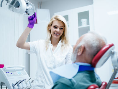
During the physical portion of the screening, the clinician performs a systematic visual inspection and gentle palpation of the head, neck and oral tissues. They look for warning signs such as red or white patches, lumps, persistent sores that do not heal, areas of induration (firmness), unexplained bleeding, or sudden changes in speech, swallowing or sensation. Documentation of any findings, including photographs in some offices, helps track change over time.
Many practices use adjunctive technologies to aid detection, such as specialized lights, vital dyes, or fluorescence devices. These tools can highlight areas that merit closer attention, but they are not substitutes for clinical judgment and are not diagnostic on their own. If a suspicious lesion is identified, the next step is referral for a definitive evaluation, which may include biopsy and pathology testing.
Patients should feel empowered to report symptoms between visits. Signs that warrant prompt attention include a non‑healing sore, a new lump in the neck, persistent throat pain, difficulty swallowing, or numbness that does not have an obvious cause. Early communication with your dental provider speeds evaluation and, when necessary, referral for further care.
Catching oral cancer at an early stage broadens treatment choices and generally improves prognosis. Smaller, well‑localized tumors often require less extensive surgery or radiation and are associated with better functional and cosmetic outcomes. Conversely, advanced cancers can necessitate more aggressive treatment and carry higher risks of long‑term effects.
Routine screening contributes to earlier detection by identifying abnormalities before they become symptomatic or visibly extensive. For people with elevated risk factors, closer monitoring can identify subtle changes sooner, allowing clinicians to act quickly. Regular screenings also create the opportunity for prevention-focused conversations about risk reduction, such as tobacco cessation and HPV awareness.
Although no screening program guarantees prevention, combining regular clinical checks with timely follow-up for suspicious findings is the most effective strategy we have to reduce the personal and societal impact of oral cancer. In short, screening saves time, reduces uncertainty and, most importantly, can save lives.
At Vernon Woods Dental & Implant Center our team follows a clear, patient-centered protocol for oral cancer screening. Every new patient and every routine recall visit includes a careful assessment of risk, a focused visual and tactile exam, and documentation of any anomalies. We aim to make the process thorough yet comfortable, explaining what we are looking for and why it matters.
If we identify an area of concern, we discuss the finding with the patient and outline the most appropriate next steps. For lesions that require definitive diagnosis, we coordinate referrals to oral and maxillofacial specialists, ENT physicians, or oral pathologists for biopsy and pathology. Our role emphasizes timely communication and continuity of care so patients move efficiently from detection to diagnosis and treatment planning.
Beyond immediate evaluation, we prioritize education and prevention. Our clinicians spend time reviewing warning signs to watch for at home and advise on lifestyle changes that lower risk. We also document findings so future comparisons are straightforward, which helps the team recognize meaningful change over time.
Regular oral cancer screening is a simple, effective component of comprehensive dental care. If you have questions about what occurs during a screening or would like to learn more about the process at our Sandy Springs office, please contact us for additional information and guidance.
Early detection matters. Reach out to learn how oral cancer screening is incorporated into your routine dental care and what you can do to lower your risk.


An oral cancer screening is a focused exam that looks for early signs of cancer in the mouth and throat. The screening combines a visual inspection and gentle palpation of the lips, tongue, gums, floor of the mouth, cheeks and neck to identify unusual tissues or lumps. The goal is to find lesions or changes when they are small and more likely to respond well to treatment.
Screening is noninvasive, quick and typically performed during a routine dental visit as part of preventive care. It creates a baseline record so that clinicians can notice even subtle changes over time. When warranted, the clinician will recommend additional testing or referral for definitive diagnosis.
Everyone benefits from periodic oral cancer screening, but certain people should be monitored more closely because of higher risk. Adults with tobacco use, heavy alcohol consumption, a history of head and neck radiation, prolonged sun exposure to the lips, or known HPV exposure typically need more frequent checks. Age and medical history also influence the recommended cadence of exams.
Your dental team will tailor screening frequency to your individual risk and overall oral health. Many practices perform a screening at every routine recall visit, which may be every six months for most patients. If you have specific risk factors or a history of suspicious lesions, the clinician may recommend shorter intervals and closer monitoring.
A typical screening begins with a review of your medical and dental history and questions about symptoms such as pain, lumps, or trouble swallowing. The clinician conducts a visual inspection of oral tissues and palpates the head, neck and oral structures to detect firmness, lumps or other abnormalities. Documentation of findings and, when appropriate, intraoral photographs help track any changes over time.
Some clinicians use adjunctive aids such as special lights, adjunct dyes, or fluorescence devices to highlight areas that warrant closer attention, but these tools do not replace clinical judgment. If a suspicious area is found, the next steps could include closer observation, imaging or referral for biopsy and pathology. The dental team will explain any findings and the recommended follow-up so patients understand what to expect.
Certain signs merit prompt evaluation rather than waiting for a routine appointment, including a sore or ulcer that does not heal within two weeks, a new lump in the mouth or neck, or unexplained numbness or persistent pain. Other warning signs include persistent hoarseness, difficulty swallowing, or red and white patches that do not resolve. Sudden changes in speech or a feeling that something is caught in the throat also deserve timely assessment.
Reporting these symptoms early improves the chances of accurate diagnosis and timely treatment. If you notice any of these changes between visits, contact your dental provider to arrange an evaluation. Early communication allows clinicians to prioritize assessment and coordinate any necessary referrals quickly.
Tobacco use and heavy alcohol consumption remain two of the strongest and most well-established risk factors for oral cancer. Infection with high‑risk strains of human papillomavirus (HPV) has become a major contributor to oropharyngeal cancers and has changed the epidemiology of the disease. Other contributors include prolonged sun exposure to the lips, a history of radiation to the head or neck, chronic irritation, and certain dietary or immune factors.
While risk varies among individuals, no one is completely immune, so screening and awareness are important for everyone. A thorough medical and social history helps clinicians assess relative risk and determine an appropriate follow-up schedule. Discussing modifiable risks with your provider can also lead to targeted prevention strategies.
Several adjunctive technologies can assist clinicians in identifying areas that merit closer inspection, including tissue fluorescence devices, reflective light tools and topical dyes. These aids can make abnormal tissue more apparent during the exam, but they are not diagnostic on their own and must be interpreted in the context of the clinical exam. Clinical judgment, history and, when indicated, biopsy remain the standards for definitive diagnosis.
Adjunct tools are most useful as part of a comprehensive approach rather than a standalone test. They can be particularly helpful when tracking suspicious areas over time or when lesions are subtle. Your clinician will explain any tools used and how the results inform next steps in evaluation or management.
If a suspicious lesion is identified, your clinician will document its appearance, size and location and discuss the finding with you. The usual pathway includes closer monitoring, diagnostic imaging when indicated, or referral to an oral and maxillofacial specialist, ENT physician or oral pathologist for biopsy and pathology testing. Biopsy is the only way to obtain a definitive diagnosis and guide treatment planning.
The dental team helps coordinate timely referral and communicates with specialists to support continuity of care. While waiting for further evaluation, clinicians may give guidance on symptoms to watch for and steps to reduce irritation or infection. Clear communication and prompt follow-up are priorities to ensure appropriate and efficient diagnosis.
Early detection typically expands treatment options and is associated with better functional and cosmetic outcomes. Small, localized lesions are often treated with less extensive surgery and may require lower doses of radiation or less aggressive adjunctive therapy. This can translate into better preservation of speech, swallowing and appearance compared with treatment for advanced disease.
Detecting cancer at an earlier stage also tends to improve overall prognosis and reduces the likelihood of complex reconstructive procedures. Regular screening and rapid follow-up for suspicious findings are practical steps that increase the chance of catching disease early. In this way, routine screening plays a meaningful role in improving patient outcomes.
Risk reduction begins with modifying known behaviors such as quitting tobacco use and limiting alcohol consumption, both of which significantly lower oral cancer risk. Protecting the lips from prolonged sun exposure with sunblock and lip balm, maintaining a nutritious diet rich in fruits and vegetables, and addressing chronic reflux or irritation can also help reduce risk. For HPV‑related oropharyngeal cancer, vaccination and safer sexual practices are important preventive measures.
Regular dental visits that include oral cancer screening are another key prevention strategy because they promote early detection of changes before symptoms emerge. Discussing your individual risk factors with your dental provider allows for a personalized prevention and surveillance plan. Combining lifestyle changes with routine professional care offers the most effective approach to lowering risk.
At Vernon Woods Dental & Implant Center our team conducts a risk assessment and focused oral cancer screening at every new patient visit and routine recall appointment, documenting findings to establish a baseline for future comparison. We explain any observations to patients, provide guidance on symptoms to monitor at home, and recommend next steps when further evaluation is necessary. The practice uses clear communication and thorough documentation to make follow-up straightforward for patients.
When definitive diagnosis is required, we coordinate referrals to oral surgeons, ENT specialists or oral pathologists and share relevant records to support continuity of care. Our clinicians prioritize timely notification and clear instructions so patients understand the referral process and expected timelines. This structured approach helps ensure that suspicious findings receive appropriate and prompt evaluation.
Ready to schedule your first visit and to join the Vernon Woods Dental & Implant Center family?
Scheduling your visit or getting answers to your questions is simple. The helpful team at Vernon Woods Dental & Implant Center is here to assist you! We can easily help you book appointments, explain treatment details, and address any concerns you have. Connect with us however you prefer—by phone or our quick online form. Don't delay your smile goals; contact us today and experience the comfort and confidence that personalized dental care provides.
Back to top