
An intraoral camera is a compact, pen-sized imaging tool that captures close-up, full-color views inside the mouth. Unlike the unaided eye, this device provides magnified detail of tooth surfaces, gum margins, and other soft tissues, enabling clinicians and patients to see the same high-resolution images in real time. The clarity of these images helps surface-level conditions become obvious at a glance and makes subtle changes easier to monitor over time.
Because the camera records color and texture, it highlights early signs of enamel wear, cracks, chips, staining, and soft-tissue irritation that can otherwise be overlooked. It is particularly useful for examining hard-to-view areas—such as the back molars or the undersides of crowns—without causing discomfort from excessive probing. These images can be displayed immediately on a chairside monitor, which helps both the care team and the patient evaluate what is happening and why certain recommendations are being made.
Beyond immediate viewing, intraoral camera images often become part of a patient’s clinical record. That documentation establishes a visual baseline that clinicians can reference during follow-up visits, helping to detect progression or improvement. For patients, seeing a photographic record of their mouth makes dental findings more concrete and understandable than a verbal description alone.
High-resolution intraoral photographs complement other diagnostic tools like visual exams and radiographs by offering surface-level context. Clinicians use these images to corroborate findings—confirming that a dark spot seen on an X-ray matches an area of enamel breakdown, for example—or to reveal cosmetic issues that require a different treatment pathway. When combined with clinical probing and imaging, intraoral photos improve diagnostic confidence and reduce guesswork.
Captured images also streamline treatment planning. Detailed photographs can be reviewed alongside digital records to map a sequence of care, illustrate the need for restorative work, or guide conservative monitoring strategies. Because the pictures are easily saved, they serve as a precise reference for shaping restorations, selecting materials, and aligning the work of specialists or dental technicians involved in a patient’s care.
For complex cases, intraoral images facilitate clear communication between providers. A dentist can annotate photographs and share them with a periodontist, endodontist, or prosthodontist to convey exact visual concerns. This shared visual language speeds collaboration, helps ensure consistent clinical goals, and reduces the risk of misinterpretation when more than one clinician contributes to a treatment plan.
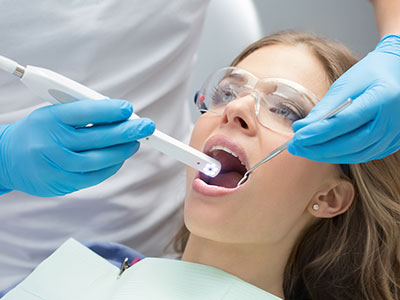
An intraoral camera exam is quick, noninvasive, and generally comfortable for patients of all ages. During a routine checkup or a focused diagnostic visit, the clinician or hygienist will gently guide the camera around the teeth and along the gumline while you sit in the dental chair. Because the device is small and handheld, it can access tight spaces without causing significant gagging or discomfort.
The procedure takes only a few minutes and is often integrated into the standard clinical exam. As images are captured, they appear on a monitor so your provider can point out areas of interest and explain what the photos show. This live review gives patients the opportunity to ask questions and helps them understand the rationale behind any recommended next steps.
Captured photos are usually labeled and stored in the patient’s digital chart so the clinical team can retrieve and compare them at future visits. This archival capability makes follow-up appointments more efficient: providers can reference previous images to determine whether a concern has progressed, stabilized, or responded to treatment, improving continuity of care.
When patients can actually see the condition of their teeth and gums, it shifts the dynamic from passive to participatory. Visual evidence supports clearer explanations, which improves informed decision-making and helps patients feel more confident about consent. Rather than relying only on technical language, a clinician can show a photo of a hairline crack or a stain and demonstrate why a particular intervention is recommended.
Intraoral images are also a powerful educational tool for at-home care. By pointing out areas where plaque accumulates or where brushing technique could improve, clinicians empower patients with specific, actionable guidance. Many patients find that seeing a photograph of a problem area motivates better oral hygiene habits and a stronger commitment to preventive measures.
Finally, visual records reduce uncertainty. Toothaches and unexplained sensitivity can be worrying; clear images help identify potential causes and set realistic expectations for follow-up tests or treatments. Knowing exactly what the clinician sees helps patients plan next steps and reduces anxiety associated with the unknown.
Intraoral photography is one component of a broader digital workflow. Images integrate smoothly with electronic health records, digital radiography, CBCT scans, and CAD/CAM systems to form a comprehensive clinical picture. When combined, these technologies produce a richer account of oral health—surface detail from the camera, internal structure from radiographs, and three-dimensional context from cone-beam imaging.
This integration improves treatment accuracy. For instance, photographs can be paired with digital impressions to guide restorative design, ensuring that a crown or veneer matches the surrounding teeth in shape and shade. Similarly, annotated images can accompany laboratory prescriptions so dental technicians have a precise visual reference for color and morphology while fabricating prosthetics or appliances.
Stored intraoral photographs also support continuity of care across providers and over time. If a patient needs a referral or seeks a second opinion, high-quality images clarify previous findings and reduce the need for repeat imaging. That continuity preserves diagnostic history and helps receiving clinicians make informed recommendations without starting from scratch.
At Vernon Woods Dental & Implant Center, intraoral imaging is part of a patient-centered approach that combines advanced tools with clear communication. By making the mouth visible in vivid detail, this technology helps clinicians provide accurate diagnoses, design thoughtful treatment plans, and involve patients meaningfully in decisions about their care.
In summary, intraoral cameras transform how oral health is examined, explained, and documented. They offer immediate, magnified views that support precise diagnosis, improved patient education, and better coordination with other dental technologies. If you have questions about how intraoral imaging fits into your care plan or would like to learn more about the technology used at our Sandy Springs practice, please contact us for more information.


An intraoral camera is a compact, handheld imaging device designed to capture high-resolution, full-color photographs inside the mouth. It typically combines a small video camera with powerful LED illumination to reveal tooth surfaces, gum margins, restorations, and other soft tissues in fine detail. Images are transmitted in real time to a chairside monitor so the clinician and patient can review findings together.
The device works by focusing light and optics on a targeted area while a digital sensor records the reflected image, producing clear photos or video clips. Many systems allow still-frame capture, annotation, and storage directly in the patient record for future comparison. Because the camera records color and texture, it highlights subtle changes that may be missed during a visual exam alone.
An intraoral camera can reveal early signs of enamel wear, hairline cracks, chips, and surface staining that are difficult to see with the unaided eye. It also captures soft-tissue conditions, such as inflammation, recession, lesions, or food packing around restorations, making these issues easier to document and explain. The magnified view helps both clinicians and patients identify focal problems and prioritize care.
Beyond immediate problems, intraoral photos establish a visual baseline that clinicians can reference during follow-up visits to track progression or improvement. This capability supports conservative monitoring when appropriate and helps determine the timing for interventions. Visual records also make it simpler to communicate specific concerns to specialists or laboratory technicians when collaborative care is needed.
Intraoral photography complements radiographs by providing surface-level context that X-rays cannot show, such as color, texture, and the exact location of external defects. While radiographs reveal internal structures and bone levels, intraoral images confirm whether a radiographic finding corresponds to an observable surface lesion or cosmetic concern. Together, these tools increase diagnostic confidence and reduce the need for speculative treatment decisions.
When paired with probing, radiographs, or three-dimensional imaging like CBCT, intraoral photos help form a more complete clinical picture for treatment planning. Clinicians can correlate an X-ray shadow with a photographed enamel defect or soft-tissue change, improving accuracy in diagnosis. This integrated approach supports more predictable restorative outcomes and clearer communication among providers.
An intraoral camera exam is typically quick, noninvasive, and comfortable for patients of all ages. The clinician or hygienist will gently guide the small camera around your teeth and along the gumline while you are seated in the dental chair, capturing a series of images that appear on a nearby monitor. Because the device is compact, it can access tight spaces without causing significant gagging or discomfort.
As images are captured, your provider will often review them with you in real time and point out areas of interest, which gives you an opportunity to ask questions and better understand recommended next steps. Captured photos are usually labeled and saved in your digital chart so the team can compare them at future visits and monitor changes over time. This process enhances transparency and helps you participate more actively in your care.
Yes, intraoral photographs are commonly saved as part of the patient’s electronic health record and serve as a visual baseline for future comparisons. Stored images allow clinicians to determine whether a lesion or restoration has changed, stabilized, or responded to treatment during subsequent visits. Archiving these photos reduces uncertainty and can eliminate the need for repeat imaging when providing continuity of care.
Saved images also facilitate communication with specialists and dental laboratories by providing precise visual references that complement clinical notes and radiographs. Annotated photographs can accompany referrals or laboratory prescriptions to convey exact concerns, shade information, or morphology preferences. This documented history improves coordination and helps ensure consistent treatment outcomes across providers.
Detailed intraoral images help clinicians map a course of care by illustrating the exact extent and location of defects, wear, or cosmetic irregularities that influence treatment choice. Photographs can be reviewed alongside digital impressions and radiographs to determine whether a restoration, a conservative repair, or continued monitoring is the best option. They also help in selecting appropriate materials and shades for crowns, veneers, or other restorations.
For restorative cases that involve laboratories or specialists, intraoral photos serve as a precise visual brief that improves fabrication accuracy and consistency. Technicians can use annotated images to match color, contour, and occlusal relationships more faithfully. As a result, prosthetics and restorations are more likely to integrate smoothly with the surrounding dentition and meet the planned esthetic goals.
Yes, intraoral cameras can detect early surface signs that often precede more advanced dental disease, such as initial enamel breakdown, early recurrent decay around restorations, and gingival irritation. Because the camera magnifies areas of interest and records color detail, clinicians may spot subtle changes that warrant preventive measures or closer surveillance. Early detection enables more conservative management and can prevent progression to more invasive treatment.
However, intraoral photography is one piece of the diagnostic puzzle and is most effective when used with probing, radiographs, and clinical examination. Some lesions or internal defects remain invisible on the surface and require radiographic or three-dimensional imaging for full assessment. Combining these modalities ensures that both surface and subsurface conditions are evaluated comprehensively.
Intraoral imaging is generally well tolerated across age groups because the cameras are small, lightweight, and noninvasive. The procedure typically takes only a few minutes and can be adapted for children, anxious patients, or those with limited mouth opening by using gentle technique and clear explanation. There is no radiation exposure from intraoral photography since it relies on visible light rather than X-rays.
Clinicians and hygienists take routine precautions to ensure hygiene and patient comfort, such as using disposable barriers on camera tips and employing gentle retraction as needed. If a patient has sensitive tissues or a strong gag reflex, the provider can modify the approach to minimize discomfort while still obtaining useful images. Overall, intraoral cameras are a safe and patient-friendly diagnostic tool.
Intraoral photographs integrate smoothly with electronic health records, digital radiography, CBCT scans, and CAD/CAM systems to create a comprehensive digital workflow. Images can be imported into treatment planning software, combined with digital impressions, or used to annotate areas of concern for laboratory communication. This interoperability enhances the precision of restorative design and improves coordination among the clinical team and external partners.
When clinical images, three-dimensional scans, and radiographs are reviewed together, clinicians gain a richer understanding of both surface anatomy and underlying structures. That fuller perspective supports more accurate diagnoses and more predictable treatment outcomes. Additionally, digital records simplify referrals and second opinions by providing receiving clinicians with a clear visual history without the need for repeat imaging.
Intraoral images transform abstract clinical findings into concrete visual evidence, helping patients understand the nature and location of dental issues. Viewing photographs on a chairside monitor allows clinicians to explain conditions in plain language while pointing to the exact area of concern, which enhances informed consent and shared decision-making. Visual feedback also makes at-home oral hygiene instruction more actionable by showing specific plaque accumulation or brushing lapses.
At Vernon Woods Dental & Implant Center in Sandy Springs, we use intraoral imaging to involve patients directly in their care and to document progress over time. Seeing photographic records often increases patient engagement, supports adherence to recommended preventive measures, and reduces uncertainty about treatment paths. This transparent approach fosters trust and helps patients take an active role in maintaining their oral health.
Ready to schedule your first visit and to join the Vernon Woods Dental & Implant Center family?
Scheduling your visit or getting answers to your questions is simple. The helpful team at Vernon Woods Dental & Implant Center is here to assist you! We can easily help you book appointments, explain treatment details, and address any concerns you have. Connect with us however you prefer—by phone or our quick online form. Don't delay your smile goals; contact us today and experience the comfort and confidence that personalized dental care provides.
Back to top