
Digital radiography replaces traditional film with electronic sensors and computer processing to capture detailed images of your teeth, gums, and jaw. Rather than waiting for film to develop, these images appear on a monitor within seconds, giving clinicians immediate visual feedback. This speed doesn’t just save time — it changes how quickly a dentist can identify issues and explain findings to a patient in real time.
Beyond convenience, digital radiography supports clearer, more consistent images. Software tools allow clinicians to adjust contrast, zoom into areas of interest, and compare images side-by-side, making subtle changes easier to spot. For patients, this translates to more confident diagnoses and treatment plans that are based on clearer visual evidence.
The move to digital also aligns with broader shifts in dental practice toward integrated, data-driven care. Digital images can be incorporated into electronic health records and used alongside other technologies — such as digital impressions and three-dimensional imaging — to provide a more complete picture of oral health.
One of the most important benefits of digital radiography is its ability to reduce radiation exposure compared with older film methods. The sensors used in digital systems are more sensitive to X-rays, so less radiation is required to produce a diagnostically useful image. This reduction is meaningful for routine care, particularly for patients who need periodic monitoring over time.
Safety is a core consideration in how modern dental offices use imaging. Clinicians follow established guidelines to minimize exposure, selecting the smallest necessary field of view and using protective measures when appropriate. The result is imaging that supports patient health while keeping radiation exposure as low as reasonably achievable.
Digital systems also eliminate the need for chemical processing, removing hazardous developer solutions from the office environment. That reduces potential staff and environmental risks, and simplifies the imaging workflow — a practical advantage that complements the safety benefits for patients.
Digital radiography provides more than just faster pictures; it delivers analytical tools that improve diagnostic accuracy. Adjustable image settings allow practitioners to highlight specific structures, detect early decay, evaluate bone levels, and assess root anatomy with greater clarity than many film-based images. These enhancements can make the difference in catching problems earlier when treatment is simpler and less invasive.
Because images are stored digitally, clinicians can readily compare current and past radiographs to track progression or healing. This historical perspective helps in monitoring conditions like periodontal disease, root resorption, or bone changes after implant placement. It also supports collaborative care when coordination with specialists is needed.
Integration with other digital modalities expands diagnostic capabilities further. Digital radiographs can be combined with cone beam CT scans, intraoral photographs, and digital impressions to create comprehensive treatment plans that consider both function and esthetics. The cohesive digital record helps clinicians plan with precision and communicate expected outcomes to patients.
Digital sensors are designed with patient comfort in mind. Many offices use thin, flexible sensors or phosphor plate systems that are easier to position and less intrusive than some traditional film holders. Faster imaging means fewer retakes and shorter appointments, reducing stress for patients who may feel anxious or uncomfortable in the chair.
From the patient’s perspective, one of the most tangible benefits is clearer communication. With images displayed on a monitor, clinicians can walk patients through what they’re seeing, annotate areas of concern, and show comparative images that illustrate change over time. This visual approach helps patients understand diagnoses and participate more confidently in treatment decisions.
Digital images also simplify coordination of care. When referrals or consultations are necessary, images can be shared securely with a specialist, lab, or another provider without the delays and degradation that can occur with physical film. That smoother exchange improves continuity of care and helps maintain treatment momentum.
Modern digital radiography systems are supported by robust protocols for storage, backup, and security. Images are archived electronically and backed up regularly to protect against data loss. Many practices use secure, encrypted systems that meet industry standards for patient privacy, ensuring that radiographic files are handled responsibly throughout their lifecycle.
Maintaining image quality and diagnostic value requires ongoing investment in staff training and equipment maintenance. Clinicians and team members receive instruction on proper sensor placement, exposure settings, and software use so images are consistently useful for diagnosis. Regular calibration and software updates keep systems performing reliably.
At Vernon Woods Dental & Implant Center, our adoption of digital radiography reflects a broader commitment to accurate, efficient, and patient-centered care. We use technology as a tool to enhance clinical judgment, improve communication, and deliver treatment decisions founded on the best available information.
In summary, digital radiography is a cornerstone of modern dental imaging: it reduces exposure, speeds diagnosis, improves patient comfort, and streamlines care coordination. If you’d like to learn more about how we use digital imaging in our practice or how it might factor into your dental care, please contact us for more information.


Digital radiography uses electronic sensors and computer processing to capture images of teeth, gums and the jaw in place of film. Unlike traditional film, images appear on a monitor within seconds, allowing clinicians to review results immediately and reduce the need for retakes. This immediate feedback supports faster diagnosis and clearer communication with patients during the appointment.
Digital systems also provide more consistent image quality because software tools let clinicians adjust contrast, zoom in on areas of interest and compare images side-by-side. Those enhancements make subtle changes easier to detect and help form treatment plans based on precise visual evidence. Digital files can be integrated into the patient record and used with other digital tools to support comprehensive care.
Digital sensors are more sensitive to X-rays than traditional film, so they require a smaller radiation dose to produce a diagnostically useful image. Because less radiation is needed for each exposure, routine imaging and periodic monitoring can be performed with a lower cumulative dose. Clinicians also select the smallest field of view and appropriate exposure settings to keep radiation as low as reasonably achievable.
Modern practices follow established safety protocols and use shielding when indicated to further reduce exposure. Eliminating chemical processing also reduces environmental and staff risks associated with developer solutions. Together, these measures make digital imaging a safer option for both patients and the clinical team.
Digital radiographs help detect a wide range of dental issues, including tooth decay, bone loss, root anatomy variations and signs of infection that may not be visible during a clinical exam. Adjustable image settings allow clinicians to emphasize specific structures and reveal subtle patterns that inform diagnosis. Comparing current images with prior studies makes it easier to track disease progression or healing over time.
Because images are stored electronically, clinicians can combine radiographs with intraoral photos, digital impressions and three-dimensional scans to form a complete diagnostic picture. This integrated approach improves accuracy when planning restorative work, endodontic treatment or periodontal care. Clear visual evidence also helps clinicians explain conditions and proposed treatments to patients.
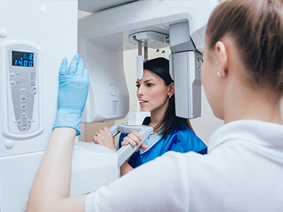
During a digital radiography appointment, the clinician or assistant will position a thin, flexible sensor or a phosphor plate in the mouth and briefly activate the X-ray unit. The process is quick and often more comfortable than traditional film holders, and images appear on a monitor within seconds for immediate review. Faster capture typically means fewer retakes and a shorter overall appointment time.
Patients can expect the clinician to review images with them, point out areas of concern and describe recommended next steps. If additional imaging is needed, such as a focused cone beam CT, the clinician will explain why it is indicated and how it complements the digital radiographs. Staff will follow standard safety measures, including the use of shielding when appropriate.
Digital radiographs provide precise information about bone levels, root positions and the relationship of teeth to critical anatomical structures, which are essential factors in planning implants and restorative work. When combined with three-dimensional imaging and digital impressions, radiographs contribute to highly accurate treatment simulations and surgical guides. This precision reduces surprises during procedures and supports predictable outcomes.
The ability to compare serial images also helps clinicians monitor healing after implant placement and assess the long-term stability of restorations. Storing images digitally facilitates collaboration with specialists and laboratories when interdisciplinary planning is required. By integrating radiographic data into the treatment workflow, clinicians can make more informed decisions and better communicate expected results to patients.
Digital radiography uses lower radiation doses than many traditional film techniques, and safety protocols are adapted for vulnerable populations such as children and pregnant patients. Clinicians only expose patients when the information gained will directly affect diagnosis or treatment, and they use the smallest appropriate field of view and protective shielding as needed. For children, exposure settings are adjusted to their size to minimize dose while maintaining diagnostic quality.
In pregnant patients, dental imaging is generally avoided unless essential for diagnosis or urgent care, and care teams discuss risks and benefits before proceeding. When imaging is necessary, strict precautions are taken to shield the abdomen and use the lowest effective exposure. Open communication with the clinician helps ensure imaging decisions prioritize patient safety.
Digital radiographs are stored electronically in secure practice management systems or imaging archives that support regular backups and redundancy to protect against data loss. Many practices implement encryption, access controls and audit logs to meet industry standards for patient privacy and secure handling of health information. Regular software updates and IT protocols help maintain system integrity and protect files throughout their lifecycle.
Staff training is a key part of reliable data management, ensuring team members understand proper file naming, storage procedures and how to securely share images when referrals are needed. The combination of technical safeguards and staff processes reduces the risk of unauthorized access and supports continuity of care. Patients can be reassured that their radiographic records are handled in accordance with professional standards.
The frequency of digital radiographs depends on each patient’s oral health, age, risk factors and the presence of symptoms rather than a one-size-fits-all schedule. Clinicians assess individual needs during exams and recommend imaging when it will provide information that influences diagnosis or treatment. For example, patients with active decay, periodontal disease or ongoing monitoring needs may require images more frequently than those with stable oral health.
Professional guidelines and best practices inform imaging intervals, but the final decision is made collaboratively between the clinician and patient based on clinical findings and medical history. Keeping a historical record of images also helps clinicians identify changes over time and avoid unnecessary repeat exposures. Open dialogue about risks, benefits and diagnostic value helps ensure imaging is used judiciously.
Yes, digital radiographs can be shared securely with specialists, laboratories and other providers to support coordinated care and timely consultations. Electronic transfer eliminates the delays and degradation that can occur with physical film, enabling precise images to accompany referrals and treatment planning. Secure file sharing and encrypted communication channels help protect patient privacy during transfers.
Sharing digital images also facilitates multidisciplinary collaboration for complex cases, such as implant planning or oral surgery, where input from specialists improves outcomes. When images are sent externally, the practice follows legal and professional requirements for patient authorization and data protection. Efficient image sharing helps maintain treatment momentum and supports comprehensive care.
Adopting digital radiography reflects a practice’s investment in modern diagnostic tools that improve accuracy, safety and patient communication. It also indicates a commitment to staff training and equipment maintenance, since achieving consistently useful images requires ongoing education and system calibration. Practices that integrate digital imaging into their workflows prioritize evidence-based care and efficient clinical decision-making.
At Vernon Woods Dental & Implant Center, our use of digital radiography supports clear diagnoses, better treatment planning and improved patient communication. By combining technology with clinical expertise, the practice aims to deliver accurate, patient-centered care and maintain high standards for imaging quality and data security.
Ready to schedule your first visit and to join the Vernon Woods Dental & Implant Center family?
Scheduling your visit or getting answers to your questions is simple. The helpful team at Vernon Woods Dental & Implant Center is here to assist you! We can easily help you book appointments, explain treatment details, and address any concerns you have. Connect with us however you prefer—by phone or our quick online form. Don't delay your smile goals; contact us today and experience the comfort and confidence that personalized dental care provides.
Back to top