At Vernon Woods Dental & Implant Center, we prioritize clear, confident diagnoses by using advanced imaging tools that reveal what standard x-rays cannot. Cone-beam computed tomography (CBCT) captures three-dimensional views of the teeth, jaws, airways, and surrounding structures, giving clinicians a comprehensive picture of your oral health. This technology helps our team evaluate complex issues with clarity and plan treatment that is safer, more predictable, and tailored to each patient’s unique anatomy.
Unlike conventional two-dimensional films, CBCT provides volumetric data that can be viewed from multiple angles and sliced into precise cross-sections. The result is a richer, more accurate dataset that supports clinical decision-making across many specialties — from implant placement to endodontic assessment and airway evaluation. In everyday practice, CBCT speeds diagnosis while helping minimize invasive exploratory procedures.
CBCT produces high-resolution, three-dimensional images that show the relationships between teeth, bone, nerves, and sinuses. Clinicians can examine root anatomy, bone volume and density, the proximity of critical structures like the inferior alveolar nerve, and subtle bony changes that may indicate pathology. These insights are essential when planning treatments where millimeters matter.
For endodontic needs, CBCT can detect hidden canals, vertical root fractures, and periapical lesions that traditional films sometimes miss. In periodontal care, the scans help assess bone loss patterns and the extent of bony defects. For orthodontic or airway evaluations, CBCT allows clinicians to visualize airway dimensions, skeletal asymmetries, and impacted teeth positions with far greater precision than planar images.
Because the images can be reformatted into 3D renderings and sectional views, the entire treatment team — including specialists when needed — can review the same dataset to arrive at a consistent, coordinated plan. This reduces uncertainty and improves communication among providers and with patients during treatment planning conversations.
One of the most significant benefits of CBCT is its role in implant dentistry. Accurate assessment of bone quality and quantity, the angulation of implant placement, and the relationship to nearby anatomic structures are all critical factors that CBCT clarifies. With three-dimensional data, clinicians can select the appropriate implant size, determine whether grafting is necessary, and design surgical guides that translate the plan precisely into the operatory.
CBCT-derived treatment plans reduce the guesswork involved in implant surgeries and allow for more predictable outcomes. By identifying potential obstacles in advance — such as thin cortical plates, sinus location, or nerve proximity — the practice can adopt measures that minimize risk, shorten surgery time, and support a smoother recovery for the patient.
When combined with digital planning tools, CBCT enables the creation of custom surgical guides and prosthetic workflows that improve accuracy and support immediate provisionalization strategies when clinically appropriate. This integration of imaging and digital design helps patients achieve functional, esthetic results with greater predictability.
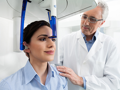
Undergoing a CBCT scan is typically quick and straightforward. The machine rotates around the patient's head while capturing a series of images that are reconstructed into a 3D volume. Most scans take less than a minute of active imaging time, and the overall visit is brief. Patients sit or stand in an open unit, making the experience comfortable and minimally intrusive for those with limited mobility or claustrophobia.
Radiation exposure from dental CBCT is greater than a single intraoral film but substantially lower than a medical CT scan. The practice follows the principle of ALARA — “as low as reasonably achievable” — selecting the smallest field of view and lowest exposure settings necessary for the diagnostic task. Our clinical team evaluates each referral to ensure a CBCT is recommended only when the expected clinical benefit justifies the imaging.
Before imaging, the clinician will review the purpose of the scan, explain positioning and safety precautions, and answer any questions. Lead precautions are used as appropriate, and the imaging protocol is customized to capture the area of interest while limiting exposure to surrounding tissues.
Acquiring high-quality images is only one part of the equation; accurate interpretation is equally critical. Our clinicians are trained to read CBCT volumes with an eye for clinically relevant findings and potential anomalies. The data are evaluated in multiple planes and window settings to identify pathology, anatomic variations, and treatment-limiting factors that can affect care.
In cases that require specialized input, the practice coordinates with radiologists or appropriate specialists for additional review. Collaborative interpretation helps ensure nothing is overlooked and supports a confident treatment plan. Clear communication of findings also helps patients understand the rationale behind recommended procedures.
We maintain protocols for record-keeping, secure image storage, and compliance with current guidelines for imaging and reporting. This systematic approach protects patient information and ensures images are available for ongoing care, consultations, or future comparisons if needed.
While CBCT is powerful, it is not a universal substitute for every diagnostic tool. Certain soft-tissue conditions and subtle mucosal changes are better assessed with other imaging modalities or clinical tests. Artifacts from metallic restorations, patient movement, and field-of-view restrictions can limit image utility in some scenarios.
Clinicians determine the appropriate imaging modality based on the clinical question at hand. For straightforward restorative checks, bitewing radiographs may suffice; for complex surgical planning, airway assessment, or unusual pathology, CBCT often provides indispensable information. The key is using CBCT judiciously — when it meaningfully improves diagnostic confidence or treatment outcomes.
Patients who have concerns about radiation exposure, pregnancy, or implant candidacy should discuss these with the care team. Together, clinician and patient can weigh the diagnostic benefits against any potential risks and choose the imaging strategy that best supports safe, effective care.
In summary, cone-beam computed tomography is a transformative diagnostic tool that enhances visualization, planning, and safety across many areas of dental care. By combining advanced imaging with experienced interpretation and a patient-centered approach, our team delivers care that is precise, thoughtful, and tailored to each individual's needs. If you would like to learn more about how CBCT may inform your treatment, please contact us for additional information.


Cone-beam computed tomography, commonly called CBCT, is a specialized dental imaging technique that creates three-dimensional views of the teeth, jaws and surrounding structures. The scanner captures a rotating series of images that are reconstructed into a volumetric dataset, allowing clinicians to view anatomy from multiple angles and in precise cross-sections. This 3D perspective reveals relationships and details that are not visible on traditional two-dimensional radiographs.
Because CBCT produces volumetric data, it supports a wide range of dental specialties including implant planning, endodontics and oral surgery. The images can be reformatted into sectional views or 3D renderings to improve diagnostic confidence and patient education. When used appropriately, CBCT streamlines diagnosis and helps clinicians develop more predictable treatment plans.
Traditional dental x-rays, such as bitewings and periapicals, provide flat, two-dimensional images that are useful for routine checks and detecting cavities. CBCT acquires volumetric data that can be sliced into axial, coronal and sagittal views, enabling accurate assessment of depth, angulation and spatial relationships. This difference is especially important when millimeter-level precision matters, such as near nerves or sinuses.
CBCT also allows clinicians to measure bone volume and density, inspect root anatomy and visualize impacted teeth in context, which two-dimensional films cannot reliably show. However, CBCT is not a replacement for all intraoral films; each modality has strengths depending on the clinical question. Clinicians choose the imaging tool that best answers the diagnostic need while minimizing patient exposure.
CBCT is typically recommended for complex diagnostic or treatment situations where three-dimensional information will change the clinical approach. Common indications include dental implant planning, evaluation of impacted or ectopic teeth, complex endodontic assessment, assessment of bony lesions and preoperative surgical planning. It is also valuable for airway assessment and evaluating craniofacial relationships in orthodontic or orthognathic cases.
For straightforward restorative checks or routine screenings, conventional radiographs often provide sufficient information and involve lower radiation. The decision to use CBCT should be individualized and based on whether the additional anatomical detail will meaningfully influence diagnosis or treatment planning. Clinicians follow established guidelines to ensure imaging is justified and focused on the region of interest.
A dental CBCT scan involves more radiation than a single intraoral film but substantially less than a medical CT scan, and modern units permit dose optimization for dental applications. Practices adhere to the ALARA principle—keeping exposure as low as reasonably achievable—by selecting the smallest field of view and lowest appropriate exposure settings for the diagnostic task. Device selection, imaging protocols and operator technique all contribute to minimizing dose while achieving diagnostic image quality.
Patients with concerns about radiation, pregnant patients or those with specific medical conditions should discuss risks and benefits with the clinician before imaging. Where feasible, alternatives or modified protocols are considered to limit exposure, and protective measures such as thyroid shielding are used when appropriate. The care team documents the justification for imaging and ensures protocols comply with professional standards.
A CBCT appointment is typically quick and noninvasive, with most active imaging taking less than a minute once the patient is positioned. The patient will be seated or standing while the scanner rotates around the head; the clinician will provide instructions to remain still and may use aids to stabilize the head for optimal image quality. The open design of most dental CBCT units is comfortable for patients and is generally well tolerated by those with limited mobility or mild claustrophobia.
Before the scan the clinician will explain the purpose of the imaging, review safety precautions and confirm the correct area to be captured. After the scan the images are reconstructed into a 3D dataset and reviewed by the clinician, who will discuss findings and next steps with the patient. If specialist interpretation is required, the images can be shared securely with consultants for collaborative review.
CBCT provides precise measurements of bone height, width and angulation that are essential for selecting implant size and position while avoiding vital structures such as the inferior alveolar nerve and maxillary sinuses. This information helps determine whether grafting is needed and supports the design of surgical guides that translate virtual plans into accurate clinical placement. The resulting predictability reduces intraoperative surprises and can shorten procedure times.
When combined with digital planning software, CBCT datasets enable prosthetically driven implant placement and coordinated restorative workflows. Virtual implant planning supports patient-specific surgical guides and, when clinically indicated, immediate provisionalization strategies. These integrated approaches enhance functional and esthetic outcomes while prioritizing patient safety.
Interpreting CBCT volumes requires review in multiple planes and window settings to identify anatomic variations, pathology and treatment-limiting factors. Clinicians trained in reading CBCT evaluate the dataset for root morphology, bony defects, proximity to nerves, sinus anatomy and any incidental findings that may affect care. Clear, systematic interpretation is important because CBCT can reveal findings beyond the initial diagnostic question.
For complex or unclear cases, the practice may collaborate with oral and maxillofacial radiologists or relevant specialists for a second opinion. This multidisciplinary review helps ensure nothing is overlooked and supports confident treatment planning. The practice maintains secure storage and documentation of images so they are available for ongoing care and consultation.
CBCT images can be affected by metal artifacts from restorations or dental hardware, and patient movement during acquisition can reduce diagnostic quality. Soft-tissue contrast is limited compared with medical CT or MRI, so certain mucosal or soft-tissue conditions may not be well visualized on CBCT. Field-of-view restrictions mean a single scan may not capture distant anatomy outside the selected region.
Because of these limitations, clinicians weigh the trade-offs between image detail, coverage and radiation dose when selecting a protocol. In some situations, alternative imaging modalities or adjunctive tests provide complementary information. Appropriate image selection and quality control are essential to obtain useful diagnostic information while minimizing repeat exposures.
CBCT allows visualization of upper airway anatomy and can provide measurements of airway volume and cross-sectional areas that are useful as part of a comprehensive airway assessment. These images help clinicians observe structural contributors to airway narrowing such as maxillary or mandibular position, enlarged soft tissues and the relationship of the tongue and palate to the airway space. Such information can inform referrals and adjunctive treatment planning for patients with suspected sleep-disordered breathing.
It is important to note that CBCT findings alone do not diagnose obstructive sleep apnea, which requires sleep studies and clinical correlation. CBCT is best used as one component of a multidisciplinary evaluation that includes medical history, physical examination and, when indicated, sleep testing. The imaging data can still be valuable for planning oral appliance therapy or surgical interventions when coordinated with a sleep medicine provider.
At Vernon Woods Dental & Implant Center, CBCT is incorporated into care when three-dimensional anatomy will improve diagnostic certainty or influence the treatment approach. The team uses focused imaging protocols, multiplanar review and collaborative interpretation with specialists as needed to ensure comprehensive evaluation and to reduce intraoperative risks. This workflow supports precise implant placement, targeted surgical planning and clearer communication with patients about recommended care.
The practice follows protocols for image justification, secure storage and documentation to maintain continuity of care and regulatory compliance. By combining advanced imaging with experienced clinical judgment, the team aims to deliver treatment that is evidence-based, predictable and centered on patient safety. Patients are encouraged to discuss any concerns about imaging so the clinician can explain the expected benefits and safeguards for their individual case.
Ready to schedule your first visit and to join the Vernon Woods Dental & Implant Center family?
Scheduling your visit or getting answers to your questions is simple. The helpful team at Vernon Woods Dental & Implant Center is here to assist you! We can easily help you book appointments, explain treatment details, and address any concerns you have. Connect with us however you prefer—by phone or our quick online form. Don't delay your smile goals; contact us today and experience the comfort and confidence that personalized dental care provides.

Back to top
